Understanding the Essential Brain Structures for Consciousness
Written on
Chapter 1: The Influence of Brain Structures on Consciousness
The intricate relationship between brain structures and consciousness is a fascinating area of study. The focus of this discussion is on identifying which specific brain regions are critical for consciousness. In simpler terms, which areas, if compromised, could cause a conscious being to lose their awareness?
The insights provided here are largely derived from lesion studies, particularly those detailed in “Neuropsychology of Consciousness: Some History and a Few New Trends” by Giovanni Berlucchi and Carlo Alberto Marzi. They emphasize a crucial point:
“Efforts to pinpoint a specific hub for consciousness based on lesions or dysfunctions that induce unconsciousness may be misguided, as consciousness is more accurately viewed as a global function of the brain's activity.”
Is There Anyone Home?
Damage to the brain stem, thalamus, or extensive regions of the cerebral cortex can lead to disruptions in consciousness. For instance, if the brain stem is injured, a person may enter a comatose state—essentially, the lights are off, yet they may still be present internally. Conversely, damage to the thalamus or substantial areas of the cerebral cortex can result in a loss of self-awareness, even if the person is physically awake. In this case, while the lights may be on, no one is home.
Thus, it becomes evident that the brain stem, thalamus, and cerebral cortex are fundamental components in the emergence of consciousness. Other lesions may disrupt self-awareness in specific ways without leading to a complete failure of consciousness. For example, damage to the hippocampus can create a unique consciousness deficit, where individuals possess a sense of self but are confined to the present moment. They understand the concept of time but cannot mentally traverse their past or envision their future.
“In summary, patients with amnesia due to hippocampal damage seem to have a factual understanding of time—where the present is preceded by the past and succeeded by the future—but lack the ability to mentally navigate through it, as they cannot recall personal experiences or imagine future scenarios.” - Berlucchi and Marzi.
Berlucchi and Marzi also note that neuropsychology often prioritizes these specific, albeit less severe, consciousness and cognitive deficits over more pronounced disruptions.
Turning the Lights On
In a conscious brain, specialized neurons within the brain stem act as an initial alert system for significant sensory information detected by our senses. Their activation can be likened to flipping a master switch, sending energizing electrochemical signals throughout the brain.
The anatomical structure known as the “ascending reticular activating system” (ARAS) is still under investigation in the human brain (see the technical notes at the end of this discussion). It is clear that there are groups of neurotransmitter-specific neurons, with their cell bodies organized into “nuclei” in the brain stem. These include serotonergic, cholinergic, and noradrenergic nuclei, which release serotonin, acetylcholine, and noradrenaline, respectively. These nuclei project their signals upward through the thalamus and/or hypothalamus into the cerebral cortex.
Incoming sensory information can activate certain subsets of brain stem neurons, leading to the release of neurotransmitters that energize the brain. The intensity of this ‘pay attention to this!’ signal can vary from a gentle nudge to a more forceful alert, depending on the nature of the sensory input.
As mentioned in “Neuropsychology of Consciousness: Some History and a Few New Trends,” multiple arousal systems operate in parallel, each responding to different motivational and emotional states. All these systems modulate activity in the thalamus and cortex, remaining active during wakefulness but quiet during sleep.
The brain stem is essential for consciousness; while it alone cannot generate consciousness, it serves as a necessary foundation.
“The renowned neurosurgeon Penfield (1978) stated that ‘to assume that consciousness or the mind has a specific localization is a misunderstanding of neurophysiology’ (page 109). However, he also theorized that a centrencephalic system, largely coinciding with the upper brain stem and hypothalamus, contains the nervous mechanisms essential for intellectual activity and the initiation of planned actions by conscious individuals” - Berlucchi and Marzi.
A Visual Representation of the Ascending Reticular Activating System
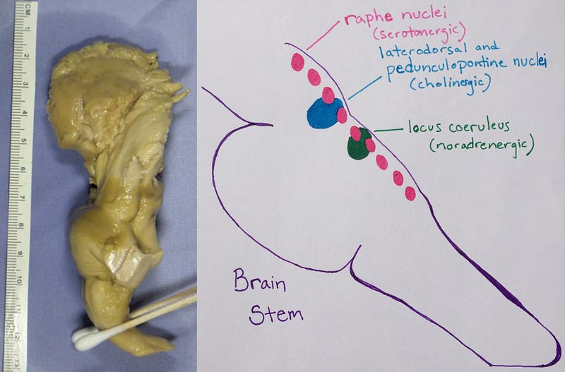
Left: Brain stem from a 78-year-old human donor, left view. From “Imaging white matter in human brainstem.” It's remarkable how this tissue is actually organized into nuclei and axon tracts. Right: Illustration by Georgeann Sack showing the location of various neurotransmitter-specific nuclei crucial for brain arousal, all located in the tegmentum of the pons and midbrain.
Reciprocal Connectivity Loops and Consciousness
Penfield, Berlucchi, and Marzi concur that consciousness is best understood as a function of the entire brain. To grasp consciousness, one must consider the dynamic processes that contribute to it. The brain operates as an organ; therefore, its metabolism is one of the key dynamic processes related to consciousness. Moreover, the brain is part of a larger organism, and the flow of information extends beyond the brain itself, interacting with the body as an open system that exchanges energy and information with the environment.
One significant dynamic process within the brain appears to be the reciprocal connectivity loops between the thalamus and cortex. These include excitatory projections from the thalamus to the cortex and vice versa. While there are additional connectivity loops between the thalamus and other brain regions, only disruptions in thalamo-cortical loops result in a loss of self-awareness. The cerebral cortex is also densely interconnected within these reciprocal loops.
As highlighted in “Mountains and Minds,” neurons in the human brain oscillate between active and inactive states. The reciprocal connectivity between the cortex and thalamus is believed to drive these oscillations.
“The reentrant architecture of vertebrate brains can also produce spontaneous rhythmic activity. The mutual exchange of action potentials transmitted through reciprocal pathways generates oscillatory behavior, similar to that observed in electrical signals recorded from active brains” - From “Reentry: A Key Mechanism for Integration of Brain Function” by Gerald Edelman and Joseph Gally.
The role of this reentrant connectivity in consciousness is a vast subject of ongoing research, which will be revisited in future discussions.
Emerging Trends in Neuropsychology
The field of neuropsychology, along with cognitive and systems neuroscience, is increasingly adopting neural stimulation techniques to deepen their understanding of consciousness—encompassing perception, attention, memory, language, and emotion. It’s worth noting that Berlucchi and Marzi clarify that neuropsychology does not primarily focus on studying consciousness itself.
Stimulation techniques, such as non-invasive transcranial magnetic stimulation and invasive electrodes used during surgical procedures, have shown promise in mapping the functions of different brain regions in living humans. For further reading, check out these two studies on using stimulation to disrupt consciousness (1, 2).
Technical Notes for Enthusiasts
In addition to the brain stem nuclei discussed, there is evidence for glutamatergic, histaminergic, and peptidergic nuclei within the hypothalamus that contribute to brain arousal initiation and regulation. Some pedunculopontine nuclei are also glutamatergic.
Each nucleus has a specific name, which can vary between research labs. Here are some nuclei associated with the ascending reticular activating system: - Locus coeruleus - Raphe nuclei - Laterodorsal and pedunculopontine nuclei - Tuberomammillary nucleus - Supramammillary nucleus - Orexin-producing neurons in the lateral and posterior hypothalamus
Axon tracts also have specific names, often named after the researcher conducting the experiments, which leads to inconsistencies across labs. Here are some tracts related to the ascending reticular activating system: - Ventral tegmental tract (rostral and caudal) - Dorsal tegmental tract (lateral and medial) - Dorsal longitudinal fasciculus - Medial longitudinal fasciculus - Superior cerebellar peduncle - Nigrostriatal tract - Medial forebrain tract
To truly understand human brain connectivity, it is essential to label specific nuclei or neuronal subtypes and conduct whole-brain imaging of long-range connections. Unfortunately, this is challenging with human tissue. Until we can label specific neuronal subtypes in living humans, visualizing cell bodies and projections of neurotransmitter-specific nuclei remains elusive. Current imaging technologies lack the resolution necessary to detect small tracts or branches from larger fiber bundles.
For a glimpse of the challenges involved, refer to “Neuroanatomic Connectivity of the Human Ascending Arousal System Critical to Consciousness and Its Disorders” and “Challenges and Opportunities for Brainstem Neuroimaging with Ultrahigh Field MRI.”
One of the most complex aspects is determining where the ascending axons terminate in the brain. As of now, no one has successfully labeled neurotransmitter-specific cell populations in the human brain stem while imaging all their targets in different brain regions. This has been accomplished in rodent models, but we should not assume that human brain anatomy follows the same patterns.
Using diffusion tensor tractography imaging (DTI) or high angular resolution diffusion imaging (HARDI), scientists can visualize axon tracts in unlabeled tissue from both living and deceased human brains. By performing histology on deceased brains and/or using human brain atlases to approximate the location of each nucleus, researchers can trace axon tracts from predetermined target regions, giving an overview of axonal pathways.
While DTI is a valuable research tool with clinical implications, it has limitations in differentiating between neuron subtypes or afferent and efferent axons. It can only indicate where bundles of axons are located but cannot reveal their directional pathways. Like many forms of brain imaging, DTI is both a science and an art form that requires years of expertise to master.
We know that neurons in the ascending reticular activating system project upwards through the thalamus into the cerebral cortex. However, the specifics of their termination remain unclear. Does a large axon bundle branch out to innervate the entire cortex, as depicted in most diagrams? Or is the innervation more targeted? Which cortical areas and layers receive these activating neurons, and near which cell types do they terminate? Is the release of neurotransmitters diffuse or highly specific? I eagerly anticipate the day we can visualize specific neurotransmitters traversing the extracellular space, as this will shed light on large-scale dynamic processes in the brain.
Understanding brain anatomy is a complex endeavor. The details of brain structure have been identified at different times, depending on the technology available, resulting in a patchwork of information. This underscores the importance of generating new atlases—new standards for the field. An interactive atlas that lists nuclei and their neurotransmitters, allowing users to visualize projections upon clicking on the nuclei, would be an incredible tool.
Acknowledgments
Special thanks to Yu Zhang, the first author of “Diffusion Tensor Tractography of Brainstem Fibers and Its Application in Pain,” and a radiologist with over 15 years of experience as a DTI tractographer, for addressing my questions regarding DTI and the ascending reticular activating system.
This video titled "Why and How Consciousness Arises" explores the fundamental questions surrounding the emergence of consciousness and its underlying mechanisms.
In this video, "The Neuroscience of Consciousness," experts delve into the intricate neural pathways and processes that contribute to conscious experience.